Cerebral Hemorrhage And Subdural Hematoma
The dura mater and the cobweb are two of the three meninges in the human brain. Meninges are structures that frame the central nervous system. Cerebral hemorrhage and subdural hematoma refer to hemorrhages in each of these two parts.
Cerebral hemorrhage and subdural hematoma: The meninges
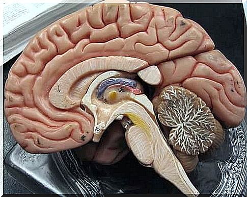
The skull and spine protect the brain and spinal cord. However, they also have another system of protection: the meninges. These actually also help with the development of nerves.
As the NIH Cancer Dictionary states: “The meninges are the three layers of tissue that cover and protect the brain and spinal cord.” We have three of these teams, which are:
- Dura mater
- Spider web membrane
- Pia mater
The outermost and thickest layer, the dura mater, is separated from the bone by the epidural space. This space does not exist in the skull since the dura mater is attached to the bone. However, it exists in the spinal cord where the blood vessels and fat are.
Beneath the dura mater lies the cobweb membrane, which is separated by the subdural space. This space occurs only when it begins to bleed and the blood separates the two meninges.
The cobweb extends through this space to the pia feeder. The subarachnoid space has cerebrospinal fluid. It actually dampens the pressure changes from sudden blows or movements.
Pia mater sticks to the nerve tissue, even in the cavities. In addition, researchers are still working to find out if there is anything inside the tissue.
Cerebral hemorrhage and subdural hematoma
In meningeal hemorrhage and subdural hematoma, blood first flows out of the blood vessels. These guys are located between the meninges. Therefore, this damages the brain tissue.
Depending on the type of bleeding , however, they will have different effects and need different treatments.
Subdural hematoma
Subdural hematoma is where blood collects in the space between the dura mater and the cobweb. This blood usually comes from blood vessels after trauma. However, there are three types of hematoma depending on how long it takes for it to appear:
- Acute subdural hematoma
- Subacute subdural hematoma
- Chronic subdural hematoma
Acute subdural hematoma
This type appears at the earliest. It is usually due to severe trauma that creates cracks in the veins that run from the cerebral cortex to the meninges.
Anyone with this usually ends up in a coma immediately. In addition, some parts of the brain stop working. Some examples of this are:
- Hemiparesis: Partial difficulty moving. This is because the injury happened to the part of the brain that handles motor skills.
- Mydriasis: The pupils become larger. This is because the injury occurred in the area that controls the iris muscle.
Subacute subdural hematoma
This appears later and is usually less severe. This is because there is less blood. In addition, the blood also begins to coagulate. However, it can also be caused by trauma.
In general, the person loses consciousness and recovers. The person will feel dizzy for several days and have trouble focusing.
Chronic subdural hematoma
This is a consequence of several injuries over time. These have small blood leaks from blood vessels. When they are not absorbed again, they end up with a hematoma. This is actually common among the elderly.
The first symptom is usually a headache, and then changes in behavior and mood. This happens little by little. In addition, people begin to sleep more, have difficulty thinking and other symptoms.
Cerebral hemorrhage
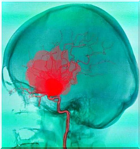
Cerebral hemorrhage is when the blood collects between the cobweb and the pia feeder. The blood usually comes from arteries and it can have many different causes. The most common is a ruptured aneurysm. However, it can also have other causes.
Aneurysms can come with headaches or even epileptic seizures before they rupture. In up to a third of cases, the trigger may be something physical with an emotional component. In addition, being in the sun for too long can happen.

The bleeding begins when they rupture. This usually occurs in people between the ages of 40 and 60 and is associated with the following symptoms:
- Vomiting
- Unfocused thoughts
- Intense headache
- Pain or discomfort from light
About 48 hours later , the symptoms of meningitis appear since the meninges are irritated. Furthermore, the neck also stiffens. In addition, you may experience eye problems, such as paralysis of eye movements.
Meningeal hemorrhages cause sequelae in up to 60% of cases. In addition, 40% of the survivors will be dependent on someone else in some way.